CAVIGEN Knowledge Hub –
3D cell culture, organoid and oxygem measurement resources
What is the CAVIGEN Knowledge Hub?
The CAVIGEN Knowledge Hub brings together scientific resources, application examples and practical guidance for researchers working with 3D cell culture, spheroids, organoids and Organ-on-Chip systems. Here you will find publications, posters, application notes, FAQs and technical information that support you in designing, running and interpreting experiments on the CaviSphere® 3D cell culture platform.
The hub is built for a growing community of labs that want to make their 3D models more predictive – from early adopters of MicroSphere and PoroSphere to advanced users of SensoSphere for oxygen measurement in 3D.
1. FAQs
Technology
Microcavities are defined, micrometre-scale wells formed in thin polymer films by microthermoforming. They act as individual “niches” in which cells aggregate and grow into 3D spheroids or organoids.
Because the geometry of the microcavities is controlled, they help to achieve more uniform 3D structures and to standardise 3D cell culture conditions across experiments.
MicroSphere is designed for standardised 3D spheroid and organoid culture with good microscopy access. PoroSphere adds controlled porosity for perfusion, co-culture and barrier models. SensoSphere enables optical oxygen measurement directly in the microcavities.
In more detail, MicroSphere uses non-porous microcavities with defined diameter to generate spheroids of similar size. PoroSphere is based on ion-track etched and chemically treated films to create well-defined pores for fluid flow and cell migration. SensoSphere combines these microcavities with oxygen-sensitive and reference fluorophores; the dynamic fluorescence quenching of the sensor dye by oxygen allows ratiometric read-out of pericellular O₂ levels in real time.
SensoSphere uses oxygen-sensitive fluorophores, where oxygen quenches the fluorescence signal, combined with a reference dye for ratiometric read-out. This allows you to calculate pericellular oxygen concentrations in real time.
The underlying principle is dynamic fluorescence quenching: the O₂-sensitive dye is excited at a defined wavelength, and in the absence of oxygen it emits a strong fluorescence signal. Collisions with dissolved oxygen molecules lead to a non-radiative energy transfer and reduce the fluorescence intensity. By measuring the ratio between sensor and reference fluorescence, and applying a two-point calibration, local oxygen concentrations can be derived with high sensitivity, even at low oxygen levels.
No. CaviSphere does not actively control oxygen; it does not include gas-control hardware or closed-loop regulation. SensoSphere enables you to measure oxygen profiles in 3D, not to regulate them directly.
The platform supports more physiologically relevant 3D models by combining standardised microcavity-based 3D cultures with pericellular oxygen measurement. The oxygen-sensitive fluorophores in SensoSphere respond to local oxygen concentration, but any control of oxygen (for example through incubator settings, gas mixing or perfusion conditions) remains within your existing equipment. The measurement data can, however, be used to refine and document physioxia-oriented culture conditions.
CaviSphere inserts and films are compatible with standard inverted and upright microscopes and with typical cell biology workflows. SensoSphere can be read out using optical oxygen measurement devices and fluorescence microscopes.
MicroSphere and PoroSphere support brightfield and fluorescence imaging and can be combined with standard plate readers for endpoint assays. SensoSphere sensor arrays have been validated in combination with commercial oxygen measurement systems (for example from PreSens) and with confocal and widefield fluorescence microscopes. The sensor chemistry is designed to work in aqueous media over a broad pH range and typical cell culture temperatures, so that you can integrate oxygen measurements into existing imaging and read-out setups.
CaviSphere helps you to make 3D models more reproducible and to document oxygen conditions, which is important for New Approach Methodologies (NAMs) and 3R strategies.
Standardised microcavities improve control over spheroid and organoid size, and SensoSphere adds quantitative oxygen measurement at the level of the 3D culture. This combination facilitates transparent reporting of culture conditions, supports the design of physioxia-oriented protocols and can contribute to in vitro models with better predictive power compared to traditional 2D systems and hyperoxic conditions.
Products & formats
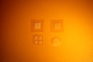
CaviSphere® is a microcavity-based 3D cell culture platform for spheroids and organoids, available as ready-to-use inserts and as films for integration into custom devices.
In more detail, all variants share a common microcavity architecture with up to 600 cavities/cm², manufactured by microthermoforming of polymer films. MicroSphere is optimised for imaging and standard assays, PoroSphere adds defined porosity for perfusion and co-culture, and SensoSphere integrates optical oxygen sensor dyes for spatially resolved oxygen measurements in 3D.
CaviSphere is available as microstructured films (“sheets”) and as pre-assembled inserts that fit into standard multiwell plates. Films are offered in different microcavity layouts and diameters, and can be cut and integrated into custom devices. Inserts combine defined microcavity fields with a convenient frame for routine handling.
The exact formats (microcavity diameters, number of cavities, materials such as PC, PS or PLA) are documented in the CaviSphere data sheets. If you are unsure which configuration fits your experiment, we recommend using our configuration tools or contacting us directly.
The optimal configuration depends on your cell type, assay read-out and culture duration. In general, smaller microcavities support smaller, more numerous spheroids, while larger cavities can host bigger organoids or more complex 3D structures.
To help you choose, we provide configuration guidance and tools that map typical applications (e.g. toxicology, Organ-on-Chip, oxygen measurement) to suitable MicroSphere, PoroSphere or SensoSphere formats. You can also contact us with your experimental requirements so we can suggest a starting point.
Inserts are ideal if you want a ready-to-use format that fits directly into standard multiwell plates and requires minimal engineering effort. Films are the better choice if you are developing custom devices or Organ-on-Chip systems.
CaviSphere inserts (e.g. 4-well inserts for 12-well plates) are pre-assembled units that can be handled like other cell culture inserts and support straightforward workflows in existing incubators, microscopes and plate readers. Films are flat microstructured foils that can be cut, bonded and integrated into microfluidic chips, bespoke chambers or Organ-on-Chip cartridges, while maintaining the same microcavity geometry and, for SensoSphere, the same optical oxygen sensor chemistry.
Yes. We provide handling protocols for all CaviSphere products, covering topics such as sterilisation, coating, cell seeding, medium changes and imaging.
These protocols are available as downloads and are continuously updated based on internal work and feedback from collaborating labs. If you have specific questions or an unusual setup, you can contact us directly for support.
Application & Handling
CaviSphere products are suitable for a wide range of 3D cell culture models, including spheroids, organoids and 3D co-cultures. Typical applications include tumour spheroids, liver spheroids, stem-cell-derived organoids and barrier models.
Microcavity arrays can be used both in simple static culture (e.g. in multiwell plates) and as components in more complex microfluidic or Organ-on-Chip setups. PoroSphere variants are particularly useful when perfusion, co-culture or migration are part of the experimental design.
MicroSphere is optimised for microscopy and the generation of spheroids for long-term 3D cell culture. It is typically used for imaging-heavy assays, toxicology screens and standard 3D culture workflows where reproducible spheroid size is important.
PoroSphere is useful for working with dynamic cell cultures and co-cultures. The defined porosity supports perfusion, barrier formation and cell migration, making it attractive for Organ-on-Chip developers and advanced in vitro models with flow.
Yes, it is possible to cultivate enough cells in CaviSphere microcavity arrays to perform PCR-based analyses.
Depending on the cell type, seeding density and culture duration, microcavity arrays can generate a sufficient total cell number for downstream molecular assays. The exact yield will depend on your specific setup, but PCR is a realistic endpoint for many 3D culture configurations.
The optimal cell number depends on your cell type and the planned culture duration, but as a starting point we generally recommend seeding around 1×10⁵ cells per sheet/insert for an initial test.
This seeding density has proven useful in first experiments with hepatoma cell lines and similar adherent cells. You can then adjust the cell number up or down based on observed spheroid size, viability and assay requirements.
Yes, staining of spheroids generated in CaviSphere microcavities is possible and commonly used.
Live/dead staining, nuclear dyes and other fluorescent markers can be applied directly in the cavities. Staining within the microcavities avoids additional handling steps and preserves the spatial context of the 3D structures for imaging.
Compared to 2D cultures, staining in 3D spheroids requires more time for the dyes to penetrate into the core of the aggregate.
In practice, the incubation time of the staining solution should be extended. We recommend up to 24 hours to ensure that even larger spheroids are homogeneously stained, especially when using nuclear or viability dyes.
There is no fixed limit to culture time in CaviSphere microcavities; it mainly depends on the number of cells seeded, their proliferation behaviour and the nutrient supply in your system.
In static cultures, medium changes and appropriate seeding densities help maintain viable spheroids over several days to weeks. In perfused systems or Organ-on-Chip setups, longer culture periods may be possible, as long as oxygen and nutrients remain within a suitable range for your cell type.
Choose inserts when you want a straightforward, plate-based 3D culture setup with minimal integration effort, for example for toxicology, imaging or routine assays. Choose films when you are incorporating CaviSphere into a custom device, microfluidic system or Organ-on-Chip platform.
Both formats support spheroid and organoid formation; PoroSphere films add porosity for perfusion and co-culture, and SensoSphere films enable integrated oxygen measurement in 3D. Inserts are often the best entry point for first experiments, while films offer maximum flexibility for advanced users.
2. Publications
Our platform builds on more than 25 years of work on microstructured 3D cell culture systems at Karlsruhe Institute of Technology (KIT).
Below you find selected research articles, a review and an expert feature authored by members of the CAVIGEN team and collaborating researchers.
Research articles
O₂-sensitive microcavity arrays: A new platform for oxygen measurements in 3D cell cultures
Grün C, Pfeifer J, Liebsch G, Gottwald E.
Frontiers in Bioengineering and Biotechnology (2023), 11:1111316.
This article introduces oxygen-sensitive microcavity arrays that combine 3D spheroid culture with optical oxygen sensing. It describes the microthermoforming process used to generate microcavity arrays from oxygen-sensitive polymer films, and demonstrates label-free, real-time oxygen measurements in the pericellular microenvironment of individual spheroids. The study also shows how 3D mitochondrial stress tests can be performed in these arrays to characterise mitochondrial respiration in 3D.
(Read article on Frontiers – DOI: 10.3389/fbioe.2023.1111316)
Physiological oxygen measurements in vitro – Schrödinger’s cat in 3D cell biology
Gottwald E, Grün C, Nies C, Liebsch G.
Frontiers in Bioengineering and Biotechnology (2023), 11:1218957.
This perspective paper discusses why dissolved oxygen concentration is a critical, yet often neglected parameter in 3D cell culture and organoid models. It reviews historical and current methods for oxygen measurement, highlights the limitations of hyperoxic “normoxia” in conventional cell culture and argues for tissue-specific physioxia. The article outlines how integrated optical oxygen sensors and microcavity-based 3D systems can enable more realistic in vitro oxygen conditions and support New Approach Methodologies (NAMs).
(Read article on Frontiers – DOI: 10.3389/fbioe.2023.1218957)
Review article
Advanced 3D Cell Culture Techniques in Micro-Bioreactors, Part I: A Systematic Analysis of the Literature Published between 2000 and 2020
Grün C, Altmann B, Gottwald E.
Processes (2020), 8(12), 1656.
This systematic review analyses 3D cell culture in micro-bioreactors over a 20-year period. It classifies different 3D culture configurations by complexity level, bioreactor design and flow mode (perfusion vs. superfusion), and surveys materials, cell types and applications. The review provides a broad context for microcavity-based 3D cell culture systems and highlights trends towards more complex co-cultures, microfluidic platforms and organ-on-chip technologies.
(Read article on MDPI – DOI: 10.3390/pr8121656)
Expert article and feature
Sauerstoffsensitive Zellkultursysteme: Organoidkultur trifft Sensorarrays
(Article in German)
This expert article on Wiley Analytical Science introduces oxygen-sensitive cell culture systems and explains how sensor arrays can be combined with organoid and 3D cell culture models. It covers the basic principles of optical oxygen sensing, discusses the integration of sensor foils and microcavities into cell culture systems and presents example applications in organoid culture and in vitro toxicology. The article is aimed at researchers interested in bringing oxygen as a controlled parameter into their 3D cell culture practice.
3. Data sheets and downloads
Here you can download key documents with technical background information on the CaviSphere® platform and its oxygen-sensing capabilities.
CaviSphere Sheets – formats and specifications
This data sheet gives an overview of the different CaviSphere® sheet formats for MicroSphere, PoroSphere and SensoSphere. It is intended for researchers who want to understand which sheet variants are available and how they can be used in standard inserts or custom devices.
It summarises the main geometrical and material features of the microstructured films, such as typical microcavity layouts, available diameters and the compatible polymers. The document also explains how CaviSphere sheets fit into common lab workflows, for example in combination with CellCrown™ inserts or as standalone films in custom setups.
White paper: PLA-based MicroSphere for more sustainable 3D cell culture
This white paper explores the use of PLA as a biobased material for MicroSphere microcavities and its potential contribution to more sustainable 3D cell culture workflows. It is relevant for labs that want to reduce the environmental footprint of their consumables without compromising data quality.
The document compares PLA to commonly used materials such as polycarbonate and polystyrene in terms of processing, optical clarity and cell culture performance, and discusses where PLA-based inserts are particularly suitable in routine 3D culture and organoid work.
4. Application notes and 3D cell culture use cases
How are CaviSphere® microcavity arrays used in real research projects?
The following examples highlight typical applications in toxicology, microfluidics and advanced read-out methods. They are meant as inspiration and starting points for your own setups.
3D toxicology and substance screening with oxygen-sensitive microcavity arrays
Focus: 3D toxicology assays, 3D mito stress tests, oxygen gradients
In this use case, hepatoma spheroids are generated and cultivated directly in oxygen-sensitive microcavity arrays. Aggregation and long-term culture are controlled within the cavities, while pericellular oxygen gradients can be monitored over time.
In more detail, the microcavity geometry ensures reproducible spheroid size, which is crucial for comparing responses to chemicals or drug candidates. Optical oxygen sensing in the same system enables parallel read-out of mitochondrial stress (for example via 3D mito stress tests) and oxygen consumption patterns. This combination supports a more nuanced interpretation of toxicological endpoints compared with conventional 2D assays.
(See also: Grün et al., Frontiers in Bioengineering and Biotechnology 2023, for details on oxygen-sensitive microcavity arrays.)
Custom oxygen-sensitive microchannels for microfluidic and Organ-on-Chip systems
Focus: customer-specific solutions, microfluidic integration, OoC and MPS development
CaviSphere microstructured films can be used as building blocks for oxygen-sensitive microchannels in microfluidic and Organ-on-Chip (OoC) systems. In this use case, customised layouts combine microcavities and optical oxygen sensor layers with channel structures tailored to a specific Organ-on-Chip or microphysiological system (MPS).
In more detail, the films are microthermoformed into channel-like geometries or bonded into existing chips, creating regions where oxygen profiles can be measured alongside 3D cell cultures or barrier models. This enables developers of OoCs and MPS to validate oxygen conditions within their chips, compare different perfusion strategies and design more physiologically oriented in vitro models before moving to animal studies or clinical samples.
(See also: Gottwald et al., Frontiers in Bioengineering and Biotechnology 2023, for a broader discussion of physiological oxygen measurements in vitro.)
PoroSphere in an MR-compatible bioreactor: capturing intracellular sodium in 3D cell culture
Focus: MR spectroscopy read-outs, intracellular Na⁺ as an early marker, tumour therapy research
In this use case, an MR-compatible bioreactor containing stacked porous microcavity arrays (PoroSphere by CAVIGEN) is used to culture 3D hepatoma cell aggregates during sodium magnetic resonance spectroscopy. The goal is to investigate how intracellular sodium (Na⁺) concentration can serve as an early marker of cell death and, consequently, as a tool to assess treatment response in tumour therapy at an early stage.
In more detail, HepG2 hepatoma cells are cultured as 3D constructs based on the protocol by Kleimaier et al. (Processes 2018, DOI: 10.3390/PR8101267). Using sodium MR spectroscopy in this 3D cell culture system, intracellular sodium changes can be monitored non-invasively following inhibition of the Na⁺/K⁺-ATPase. The MR-compatible bioreactor encloses and nurtures the cells with stacked PoroSphere films, which provide mechanical support, medium access and MR visibility during the measurement.
This combination of porous microcavity arrays and advanced MR spectroscopy helps bridge physics, biology and medicine: it allows researchers not only to follow relative changes in intracellular sodium over time, but also to approach absolute quantification in a physiologically more relevant 3D environment than traditional 2D monolayers.
If you are working on advanced in vitro models, MR-based functional read-outs or early-response markers in tumour therapy, we are happy to explore how PoroSphere-based 3D setups could support your research.
5. CAVIGEN Academy – training, videos and workshops
We are building the CAVIGEN Academy to make it easier for labs to get started with 3D cell culture in microcavities and oxygen measurement – and to move from first trials to robust, routine applications.
In the future, the Academy will combine on-demand learning and interactive formats:
- short training videos that explain key concepts, workflows and troubleshooting steps
- small-group workshops offered via selected training partners
- customised workshops on site at your lab, focusing on your specific cell models and read-outs
Planned topics include:
- setting up and optimising 3D spheroid and organoid cultures in MicroSphere
- designing perfusion, co-culture and barrier experiments with PoroSphere
- planning oxygen measurements with SensoSphere and interpreting oxygen profiles in 3D
- integrating CaviSphere into existing plate-based, microfluidic or Organ-on-Chip workflows
If you are interested in upcoming courses, training videos or a workshop at your site, we would be happy to talk about what you need and how we can support your team.